Musculoskeletal Nuclear Scintigraphy in the Sports Horse - Usefulness & Limitations in the day and age of CT/MRI
Species
Equine
Contact Hours
3 Hours - RACE Approved
Language
English
Discipline
Diagnostic Imaging
Orthopaedics
Sports Medicine
Toxicology & Pharmacology
Veterinary Partner
Equine


Recorded on: 18th October 2022
Panelists:
Sue Dyson MA, Vet, MB, PhD, DEO - Consultant, UK
Mathieu Spriet MS, DVM, DACVR, DECVDI - UC Davis, USA
Filip Vandenberghe DVM, AssocECVDI-LA - Equine Hospital Bosdreef, Belgium
Moderator:
Sarah Puchalski BSc, DVM, DACVR - Puchalski Equine Imaging, USA
CONTENT DESCRIPTION
Nuclear scintigraphy is a tried and tested imaging modality in horses. Has it however become obsolete in the wake of MRI & CT imaging more readily available across the equine veterinary sector?
An international panel of world-renowned specialists with decades of experience with advanced imaging will be discussing the use of nuclear scintigraphy for musculoskeletal disorders in sports horses. This lively exchange will cover key points, chiefly amongst them the utility of the technology in a world full of other diagnostic imaging choices. Other topics such as clinical indications, pitfalls with the interpretation, expectations from the results, and peculiarities associated with scanning in the equine patient will also be addressed. The emergence, indications and usefulness of Positron Emission Tomography (PET) scans will also be discussed.
.
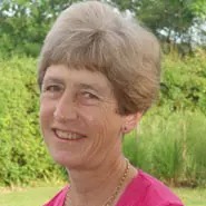
Sue Dyson qualified as a veterinarian from the University of Cambridge in 1980. After an internship at the University of Pennsylvania and a year in private equine practice in Pennsylvania, Sue returned to Great Britain to the Animal Health Trust, Newmarket. Sue ran a clinical referral service for lameness and poor performance, attracting clients from all over the United Kingdom, Ireland and continental Europe for 37 years. From 2019 she has worked as an independent consultant, combining her horsemanship skills with her veterinary experience, with the aim of maximising performance potential.
Sue’s key interests are improving the diagnosis of lameness and poor performance and maximising the opportunity for horses to fulfil their athletic potential at whatever level, taking a holistic approach to the horse, rider and tack combination, and improving approaches to diagnosis and management. She has been involved not only in providing clinical services, but also clinically relevant research and education. Sue is co-editor, with Mike Ross, of Diagnosis and Management of Lameness in the Horse and co-author of Clinical Radiology of the Horse and Equine Scintigraphy. With Sue Palmer she wrote Harmonious Horsemanship: Use of the Ridden Horse Ethogram to Optimise Potential, Partnership and Performance. She has published more than 430 papers in peer reviewed journals concerning lameness and diagnostic imaging and has lectured worldwide to veterinarians, paraprofessionals, coaches, riders and judges.
Sue is a former President of the British Equine Veterinary Association and is currently scientific advisor to the Saddle Research Trust and Moorcroft Rehabilitation Centre. Sue is also a rider, and has produced horses from novice to top national level in both eventing and show jumping. Sue holds the Instructors and Stable Managers Certificates of the British Horse Society (BHSI).
Sue has been awarded many international accolades for her work including induction into the University of Kentucky Equine Research Hall of Fame for outstanding contributions to research in equine veterinary science, Honorary Membership of the British Equine Veterinary Association and Societa Italiana Veterinari Per Equini, Italy, the American Association of Equine Practitioners Frank J. Milne Award and the Tierklinik Hochmoor Prize, Germany, for outstanding, creative and lasting work in equine veterinary medicine.
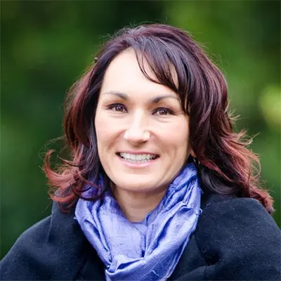
More InfoSarah Puchalski is a native of Roberts Creek, British Columbia, Canada, and grew up riding horses in the BC countryside, competing actively in 3-day eventing. She obtained a B.S. in Biology from Simon Fraser University in Burnaby, BC and went to veterinary school in Saskatoon, Saskatchewan, where she graduated with distinction in 1999. From Saskatoon, she moved to Pennsylvania as an intern/resident in field service sports medicine at the University of Pennsylvania’s New Bolton Center from 1999-2001. From New Bolton Center, Dr. Puchalski entered a four-year residency in diagnostic imaging at the University of California, Davis. She obtained board certification in the American College of Veterinary Radiology in 2004, passing the grueling examination on her first attempt.
In 2005, Dr. Puchalski joined the faculty in diagnostic imaging in the Department of Surgical and Radiological Sciences, UC Davis School of Veterinary Medicine, as an Assistant Professor. During this time, she divided her focus between didactic and clinical teaching, research, service to the school and clinical radiology. In the classroom, she led the general large animal radiology course and was a co-instructor in the equine lameness and radiology course. Her research interests predominantly surround the use of novel imaging techniques for the diagnosis of lameness conditions in equine athletes. As she has gained experience in this field, she has developed an international reputation for her expertise. Dr. Puchalski was awarded tenure at the University in 2012 and promoted to Associate Professor, and has authored scientific, educational and lay articles as well as contributed to numerous textbooks.
Dr. Puchalski left the University in late 2013 to open her own practice as a diagnostic imaging consultant. In the winter months she is based in Wellington, Florida at Palm Beach Equine Clinic and during the remainder of the year in Petaluma, California at Circle Oak Equine. As a show jumper, competing actively in Wellington, Florida at the Winter Equestrian Festival and in Northern California, she understands the challenges of equine ownership and what may be needed to keep these athletes sound.
More Info
Dr. Mathieu Spriet is a Professor of Diagnostic Imaging at the School of Veterinary Medicine at the University of California, Davis. He obtained his DVM degree from the National Veterinary School of Lyon (France) in 2002 and a Master Degree from the University of Montreal (Canada) in 2004. He has been a diplomate of both the American College of Veterinary Radiology and the European College of Veterinary Diagnostic Imaging since 2007, after completing his radiology residency at the University of Pennsylvania. Dr Spriet joined UC Davis as a faculty member in 2007. He became a diplomate of the newly created ACVR- Equine Diagnostic Imaging specialty in 2019. Dr Spriet has over 75 peer-reviewed publications (full list of publications: https://www.ncbi.nlm.nih.gov/myncbi/mathieu.spriet.1/bibliography/public/). He is a frequent speaker at national and international conferences. His main area of interest is equine musculoskeletal imaging. He has pioneered the use of positron emission tomography in horses, leading to the development of a scanner specifically designed to image standing horses. He is a consultant for advanced imaging in racehorses at several racetracks in the USA, including Santa Anita and Churchill Downs. He serves as an expert on the Racing Victoria imaging panel.
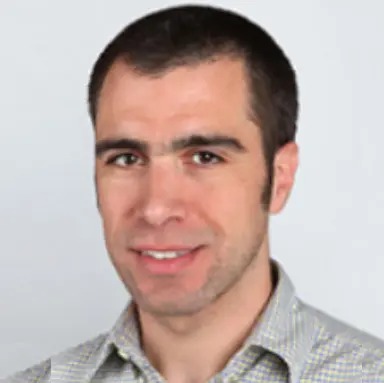
More InfoFilip graduated in 2001 and joined immediately after the orthopaedics department of the Faculty of Veterinary Medicine of Ghent University. In 2004 Filip joined the Bosdreef and became partner in 2008. Over 15 years ago Filip pioneered amongst few others in the world, on the clinical use of standing MRI in the horse. In 2011 he was awarded Associate LA ECVDI, the European College of Veterinary Diagnostic Imaging. The last years Filip has focused as well on the poor performance of the competition horse. Filip is a frequently invited speaker on national and international scientific congresses. FANC license nuclear medicine.
More InfoVeterinary Student
Online Panel Discussion
USD 20.00
Qualified Vet
Online Panel Discussion
USD 95.00
Intern/Resident/PhD (Requires proof of status)
Online Panel Discussion
USD 70.00
Vet Nurse/Vet Tech (Requires proof of status)
Online Panel Discussion
USD 70.00
If the options you are looking for are unavailable, please contact us.
No tax will be added unless you are a UK taxpayer
Choose currency at checkout


