Panel Discussion – Challenges & Opportunities in Equine Imaging
Species
Equine
Contact Hours
2 Hours - RACE Approved
Language
English
Discipline
Diagnostic Imaging
Orthopaedics
Sports Medicine
Surgery
Veterinary Partner
Equine

Time: London 7PM / Paris 8PM / New York 2PM / Sydney 4:00AM (+1)
Part of the Equine Online Diagnostic Symposium – CT, PET Imaging & Objective Gait Analysis Online Lecture Series
CONTENT DESCRIPTION
Description to follow soon.
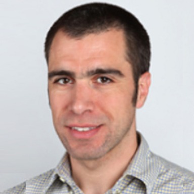
Kevin, a Missouri native, graduated from the University of Missouri College of Veterinary Medicine in 1983. He began his career in private equine practice, working in Maryland and Delaware for three years. In 1989, Kevin completed a three-year equine surgery residency and earned a Master of Science degree at the University of Illinois in Urbana-Champaign. During this time, he gained expertise in statics and dynamics, experimental stress analysis, biomedical instrumentation, and signal processing.
After finishing his surgery residency, Kevin returned to private practice near Detroit, Michigan, focusing on equine surgery and lameness. In 1990, he joined the University of Missouri as a clinical instructor and was promoted to Assistant Professor in 1991, and to Associate Professor in 1998. He is board certified by the American College of Veterinary Surgery.
Kevin's primary research interest lies in using computer-assisted gait analysis to diagnose lameness in horses. He is an active member of several professional organizations, including the Missouri Veterinary Medical Association, the American Association of Equine Practitioners, and the American Veterinary Medical Association.
More InfoDr. Mathieu Spriet is a Professor of Diagnostic Imaging at the School of Veterinary Medicine at the University of California, Davis. He obtained his DVM degree from the National Veterinary School of Lyon (France) in 2002 and a Master Degree from the University of Montreal (Canada) in 2004. He has been a diplomate of both the American College of Veterinary Radiology and the European College of Veterinary Diagnostic Imaging since 2007, after completing his radiology residency at the University of Pennsylvania. Dr Spriet joined UC Davis as a faculty member in 2007. He became a diplomate of the newly created ACVR- Equine Diagnostic Imaging specialty in 2019. Dr Spriet has over 75 peer-reviewed publications (full list of publications: https://www.ncbi.nlm.nih.gov/myncbi/mathieu.spriet.1/bibliography/public/). He is a frequent speaker at national and international conferences. His main area of interest is equine musculoskeletal imaging. He has pioneered the use of positron emission tomography in horses, leading to the development of a scanner specifically designed to image standing horses. He is a consultant for advanced imaging in racehorses at several racetracks in the USA, including Santa Anita and Churchill Downs. He serves as an expert on the Racing Victoria imaging panel.
More InfoChris is a leading researcher in equine orthopedics at the University of Melbourne, where he employs a multidisciplinary approach involving biomechanics, microstructural analysis, and epidemiology. His work integrates clinical focus, drawing on his expertise as a specialist equine surgeon at the Veterinary Teaching Hospital, where he has been since November 2004, investigating and treating lameness in horses.
Chris's journey includes specialist training at the University of Sydney, where he earned a PhD, followed by work as a specialist surgeon and scientist at the Animal Health Trust in Newmarket, England. He later ran his own referral practice and scintigraphy unit at the Newcastle Equine Centre from 1999 to 2004.
Chris has a strong track record of lecturing and publishing extensively on lameness and musculoskeletal injury prevention. His current research interests focus on equine limb function, subchondral bone, and mathematical modeling of equine limb injuries
More InfoKate earned her VMD from the University of Pennsylvania in 2012, after obtaining a degree in biochemistry from Tufts University. She then completed a rotating internship in equine medicine, surgery, and critical care at the New Bolton Center in Pennsylvania. Following her internship, Kate pursued a diagnostic imaging residency at Tufts University's Cummings School of Veterinary Medicine.
In 2016, Kate became board certified in diagnostic imaging by the American College of Veterinary Radiology (ACVR). That same year, she joined the faculty at the New Bolton Center, University of Pennsylvania, as an Assistant Professor of Diagnostic Imaging. Kate's clinical work focuses on the use of advanced imaging technologies such as CT, MRI, and a robotics-controlled cone-beam CT system designed for use with standing horses.
Kate is also involved in clinical research on the veterinary applications of rapid prototyping, including 3D printing. In her free time, she enjoys riding her Warmblood jumper, Catapult, and spending time with her husband, Zach, and their Labradors.
More InfoQualified Vet
Webinar
USD 120.00
Intern/Resident/PhD (Requires proof of status)
Webinar
USD 90.00
Vet Nurse/Vet Tech (Requires proof of status)
Webinar
USD 90.00
Veterinary Student (Requires proof of status)
Webinar
USD 20.00
If the options you are looking for are unavailable, please contact us.
No tax will be added unless you are a UK taxpayer
Choose currency at checkout

 Thu, 03 October, 2024
 07:00 pm - 09:00 pm
(Your Local Time Zone)


